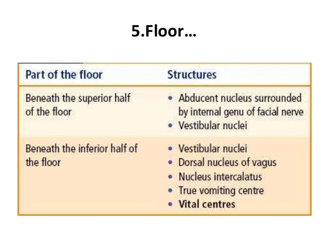
Cranial Nerve Nuclei Present In Floor Of 4th Ventricle
Cranial Nerve Nuclei Anatomy And Embryology Kenhub

Brainstem Ii Pons And Cerebellum Part 2 Cranial Nerves Skeletal System Anatomy Spinothalamic Tract

Abducens Vi Cranial Nerves Craniosacral Therapy Cranial Nerves Abducens Nerve

Mesencephalic Nucleus Google Search Cranial Nerves Pharmacology Nursing Cranial Nerves Mnemonic

Brainstem I The Medulla Organization Of The Central Nervous System Part 2 Nervous System Parts Medical Knowledge Nervous System

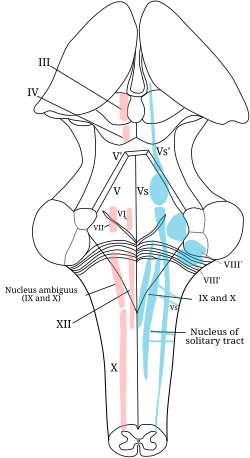
Main Motor Nucleus Nucleus Ambiguus Deep In The Reticular Formation Parasympathetic Nucleus Dorsal Nucleu Nervous System Parts Vagus Nerve Cranial Nerves

To identify patterns of cranial motor nuclei cmn displacement in cases of intramedullary brain stem tumor using neurophysiological mapping of motor nuclei on the floor of the fourth ventricle.
Cranial nerve nuclei present in floor of 4th ventricle.

Frontal Section Taken Through The Level Of The Rostral Diencephalon Where The Thala Mus Is Not Present Note Again Th Gross Anatomy Brain Anatomy Brain Parts
Hypoglossal Nucleus Wikipedia
Snp Cranial Nerve Nuclei Fa 2019 Kaplan 2018 Deja Review Hy 5th Edi Snell Flashcards Memorang

Jaypeedigital Ebook Reader
Neuroanatomy Online Lab 3 The Ventricles And Blood Supply Cranial Nerves Of The Pons

What To Expect From A Stroke Of The Medulla Oblongata Cognitive Activities Brain Stem Nursing School Notes
Https Web Duke Edu Histology Mbs Videos Neuro 1 18 20cranial 20nerve 20nuclei 20part 201 20 12 04 01 18 19 20 21 Cranial 20nerve 20nuclei Notes Pdf

Basal Ganglia Stroke Basal Ganglia Caudate Nucleus Basal Ganglia Stroke

Vestibulocochlear Viii Cranial Nerves Cranial Nerves Medical Anatomy Pharmacology Nursing
Https Web Duke Edu Anatomy Lab27 2019 2020 Lab Internal Anatomy Of The Brainstem And Spinal Cord 20 1 Pdf

Rhomboid Fossa An Overview Sciencedirect Topics
4th Ventricle 21 12 2015 Dk

Pin By Tonnianne Wisdom On System Based Medicine Frontal Lobe Corpus Callosum Brain
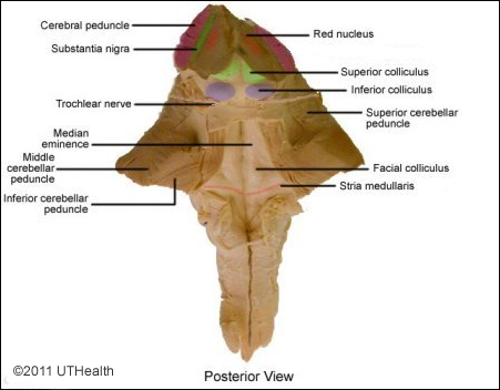
Pons Anatomy And Syndromes
Cranial Nerves Neuroanatomy

Oculomotor Nucleus Wikipedia

Clinical Neuroanatomy Twenty Eighth Edition

Cns Intro To Brain And Ventricles Medulla Oblongata Pons Mid Brain And Cerebellum Medical Knowledge Brain Anatomy Cns System
Https Encrypted Tbn0 Gstatic Com Images Q Tbn 3aand9gcqmypkl12lutfc4ut3h5nimqjgi4g Vs F3mw6n3i 6 S9d9b4 Usqp Cau

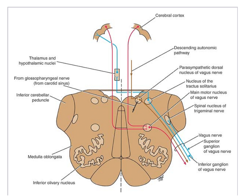
Dorsal Nucleus Of Vagus Nerve Wikipedia

Caudate Nucleus Lily And Beyond Basal Ganglia Caudate Nucleus Basal Ganglia Stroke
Abducens Nerve Wikipedia

Midsagittal Diagram Of Human Brain Brain Anatomy Human Brain Anatomy Brain Diagram
Ventricular System Of The Brain Neonatal Neurosonography Sonography Neonatal Pediatrics
Source : pinterest.com
