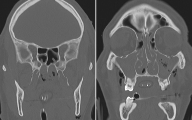
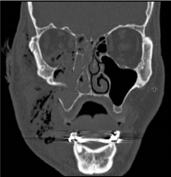
Ct Scan Obital Floor Plate

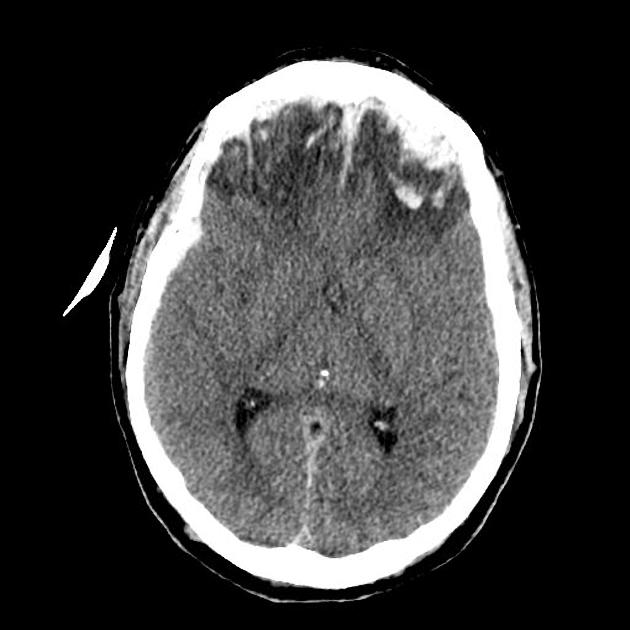
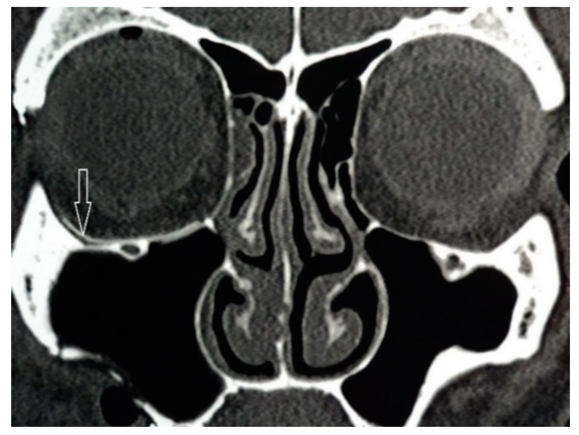
History Of Prior Surgery Bone Defect Is Seen In The Right Cribriform Plate Contrast Leak And Accumulation Within The Righ Nasal Cavity Head And Neck History

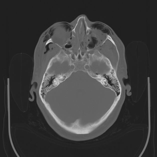
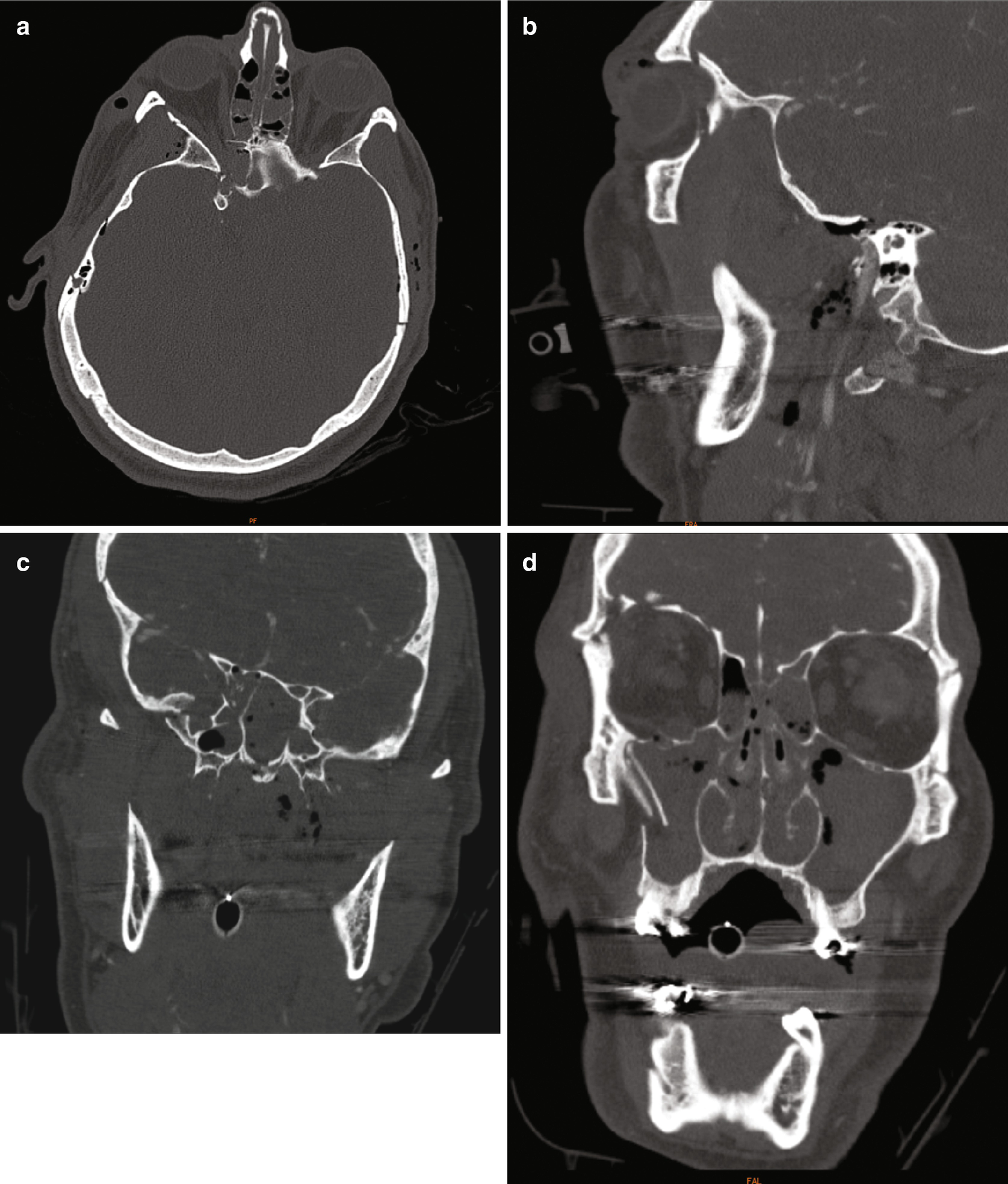
Ct Scan Of Facial Bones Axial View A Showing Fracture Of Bilateral Download Scientific Diagram

Imaging The Face Radiology Key

Orbital Blowout Fracture Radiology Reference Article Radiopaedia Org
Https Www Neiltanna Com Assets Pdf Face 18 Pdf

Face And Neck Emergencies Radiology Key
The use of a polydioxanone pds plate for orbital reconstruction was evaluated in 20 patients with various traumatic defects of the orbital floor.
Ct scan obital floor plate.
Diagnosis And Treatment Of Orbital Fractures
Https Www Zvitmedical Com Wp Content Uploads 2013 10 Doc 5 Pcl Permanent Versus Bioresorbable Implants In Orbital Floor Pdf

Intraoperative Imaging O Arm In Secondary Surgical Correction Of Post Traumatic Orbital Fractures Sciencedirect

Pin En Radiology

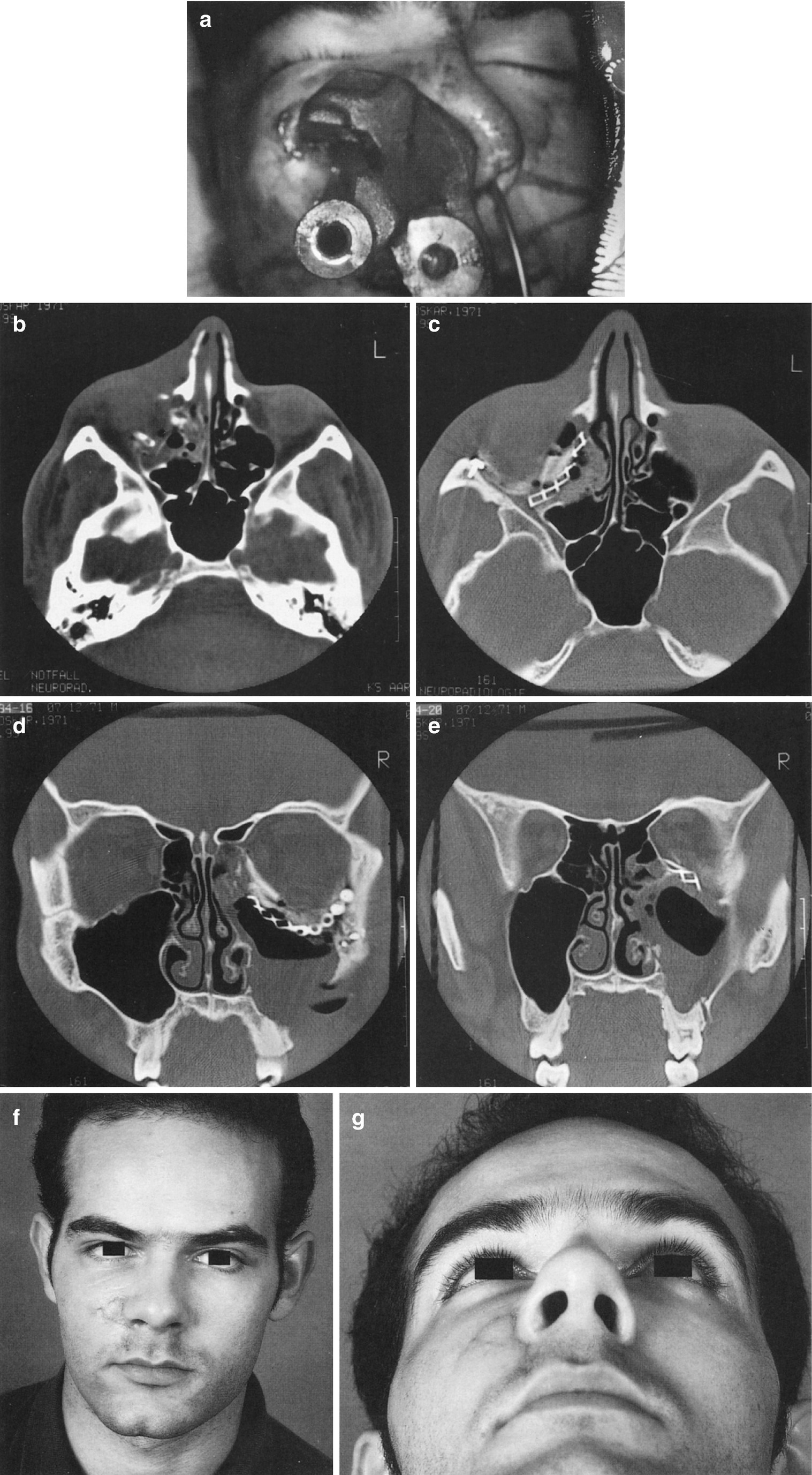
Intranasal Migration Of A 35 Year Old Orbital Plate Presenting As Unilateral Epiphora Sciencedirect
Open Reduction With Or Without Internal Fixation For Orbit Orbital Floor Fracture
Le Fort 3 Fractures Radiology Case Radiopaedia Org

Reconstruction Of Medial Wall Blowout Fracture Defect With A Combination Of Resorbable Meshed Plate And Cancellous Bone Allograft

Intraoperative Imaging Changes Management In Orbital Fracture Repair Journal Of Oral And Maxillofacial Surgery

Tips And Tricks In Surgical Management Of Maxillary Sinus Tumors Sciencedirect

Reconstruction Of A Complicated Orbital Depression Fracture With Medial Wall And Globe Repositioning In A Horse A Collaboration Across Disciplines And Specialties Mcmaster 2016 Veterinary Surgery Wiley Online Library
Orbital Reconstruction Springerlink
Le Fort Fracture Classification Radiology Reference Article Radiopaedia Org
Complications In Cranio Maxillofacial Trauma Springerlink

Radiographic Anatomy Of The Orbit And Visual Pathways Radiology Key
Https Pubs Rsna Org Doi Pdf 10 1148 Rg 2019180118
Treatment Of Zygomatic Complex Fractures With Surgical Or Nonsurgical Intervention A Retrospective Study

Lamina Papyracea Radiology Reference Article Radiopaedia Org
Https Encrypted Tbn0 Gstatic Com Images Q Tbn 3aand9gcthtq54iuk Fgkr2ypw 1wu5qgc1rdagkhlcc G9bt5uslsvnlt Usqp Cau
Jfb Free Full Text Titanium Nickelide In Midface Fractures Treatment Html

Use Of Virtual Surgical Planning And Virtual Dataset With Intraoperative Navigation To Guide Revision Of Complex Facial Fractures A Case Report Journal Of Oral And Maxillofacial Surgery

Controversies In Orbital Reconstruction I Defect Driven Orbital Reconstruction A Systematic Review Pocket Dentistry

Current And Evolving Trends In The Management Of Facial Fractures Vujcich 2018 Australian Dental Journal Wiley Online Library

Pdf Extensive Maxillary Sinus Pneumatization
Source : pinterest.com
